As often said, an ultrasound is a gynaecologist’s stethoscope. You will notice that it will be used very commonly during your antenatal visits and gives us important information about the ongoing pregnancy.
During pregnancy two ultrasounds are done as a must, one between 11 and 13 weeks and the other 18 to 22 weeks of pregnancy.
The ultrasound done between 11 and 13 weeks dates the pregnancy ie; confirms whether the date of the last menstrual period conforms to the growth of the baby or not. So, mostly it’s referred to as dating ultrasound. But it also provides important information regarding foetal formation, placental location, amniotic fluid volume, and also the competence of the cervix. Any further investigations depend on the findings of this ultrasound report.
Ultrasonography In Pregnancy
...
23/04/2024
As often said, an ultrasound is a gynaecologist’s stethoscope. You will notice that it will be used very commonly during your antenatal visits and gives us important information about the ongoing pregnancy.
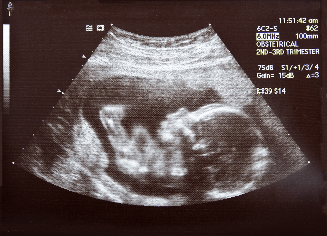
During pregnancy two ultrasounds are done as a must, one between 11 and 13 weeks and the other 18 to 22 weeks of pregnancy.
The ultrasound done between 11 and 13 weeks dates the pregnancy ie; confirms whether the date of the last menstrual period conforms to the growth of the baby or not. So, mostly it’s referred to as dating ultrasound. But it also provides important information regarding foetal formation, placental location, amniotic fluid volume, and also the competence of the cervix. Any further investigations depend on the findings of this ultrasound report.
The second ultrasound examination is planned in the second trimester between 18 and 22 weeks of gestation. The purpose of this ultrasound is more or less the same as the one in the first trimester. It confirms the dates and reviews the foetal anatomy and rules out most but not all the abnormalities.
To rule out foetal Cardio-vascular abnormalities ultrasound examination only, does not pick most of them unless they are major and have an impact on heart function. To diagnose foetal Cardiac abnormalities foetal echocardiography examination is needed. This examination is not done on all babies and is only recommended and done if there is some indication in the baby of malfunctioning like increased cardiac size or in history if such malformations are found in the siblings or other family members. If indicated the echocardiography is planned between the 26th and 28th weeks of pregnancy.
A third ultrasound examination is done between 28 and 30 weeks to diagnose some of those abnormalities that arise in the third trimester like Hydronephrosis, Ventriculomegaly, Hydrocephaly, Microcephaly, Renal agenesis, certain Gastro-intestinal abnormalities, etc.
The gender of the baby is not revealed unless the patient asks for it specifically.
Not all foetal abnormalities are picked or diagnosed on ultrasound examination.
Medicsi, established in 2006, prioritizes cutting-edge diagnostic technology in the treatment decisions of our patients. Our institution emphasizes patient comfort and employs competent senior consultants to oversee their care.
Medicsi, established in 2006, prioritizes cutting-edge diagnostic technology in the treatment decisions of our patients. Our institution emphasizes patient comfort and employs competent senior consultants to oversee their care.
We use cookies to ensure that we give you the best experience on our website. If you continue to use this site we will assume that you are happy with it.